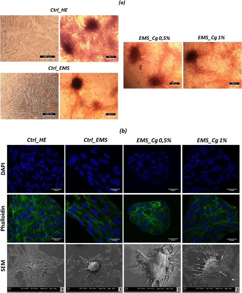
Fig. 2.
Evaluation of chondrogenic cells morphology. a Chondrogenic cultures were stained with Safranin O and observed under an inverted microscope, bar size 200 μm; magnification was set at 40-fold. b DAPI and Phalloidin labeled cells were observed using an inverted epi-fluorescent confocal microscope; scale bar size 20 μm; magnification was set at 60-fold. Cell surface and shape was assessed by the mean of scanning electron microscope; scale bar size 30 μm; images were acquired under 1000-fold magnification

