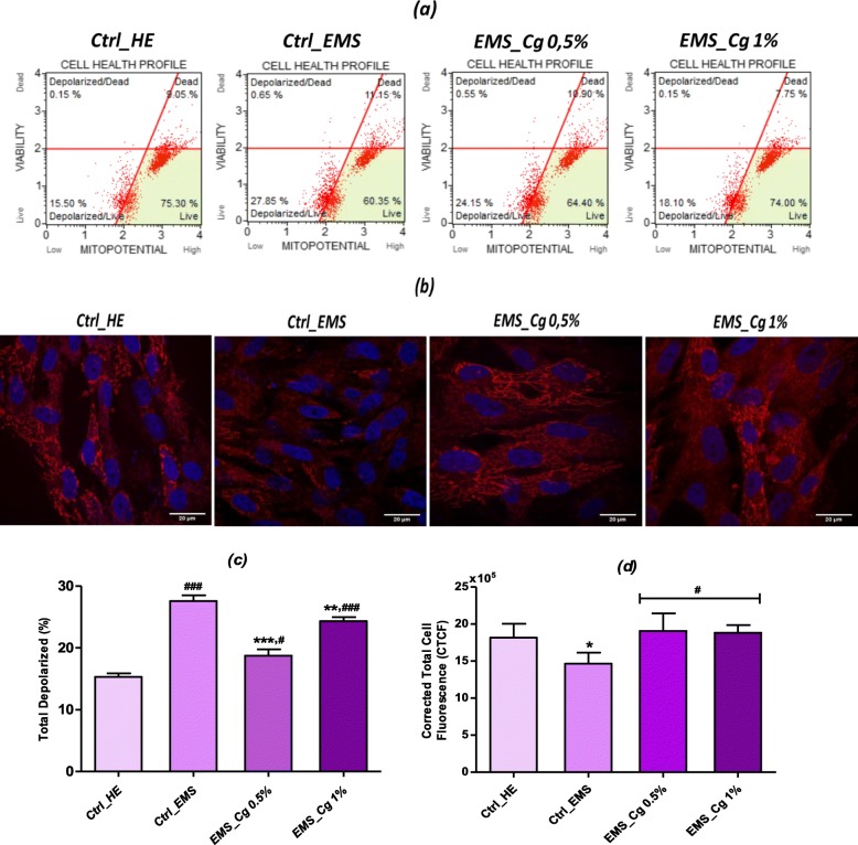
Fig. 6.
Mitochondrial membrane potential analysis. a Scattered blots representation of live and dead depolarized cells percentages for one representative experiment. b MitoRed stained cells were observed using an inverted epi-fluorescent confocal microscope; scale bar size 20 μm; magnification was set at 60-fold. c Bar charts represent the average percentages ± SD of total depolarization for three repetitions. d Bar chart representation of corrected total cell fluorescence (CTCF) for MitoRed fluorescent staining. Asterisk (*) refers to comparison of treated groups to untreated EMS cells. Hashtag (#) refers to comparison of treated groups to untreated healthy cells. */#p < 0.05, **/##p < 0.01, ***/###p < 0.001

