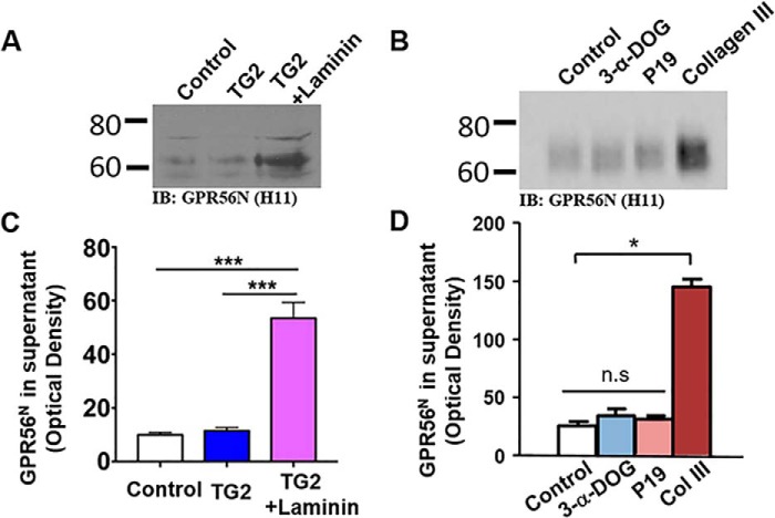
Figure 4.
Dissociation and shedding of GPR56 NTF. A and B, HEK293T Cells transfected with WT GPR56 were treated with TG2 (200 nm), TG2 plus laminin (2 μg/ml), 3-α-DOG (5 μm), P19 (20 μm), or collagen III (50 nm). Western blotting reveals the NTF of GPR56 in the supernatant. IB, immunoblot. C and D, quantification of GPR56 NTF in the supernatant. Data are presented as mean ± S.D.; n = 3; *, p < 0.05; ***, p < 0.001; ns, not significant; two-tailed Student's t test.

