Figure 3.
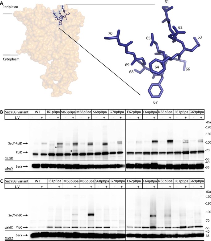
Plug domain of SecY makes multiple contacts to both YidC and PpiD. A, cartoon showing the structure of the SecYEG complex (PDB 4V6M) with the position of the plug helix (blue). Residues that were replaced with pBpa for in vivo cross-linking are indicated. B, BL21 cells expressing SecYEG variants containing pBpa at different positions of the plug domain were UV-exposed when indicated, and SecYEG was purified via the C-terminal His-tag on SecY. The purified samples were further analyzed by SDS-PAGE and Western blotting for identification of cross-linking products. The membrane was decorated with α-PpiD antibodies, and the SecY–PpiD cross-linking products as well as the co-purifying PpiD are indicated. The lower part of the membrane was also decorated with α-SecY antibodies for controlling that comparable amounts of SecY were loaded. However, because of the low quality of the α-SecY antibody, detection required longer exposure times. The SecY(I61pBpa) cross-linking product at ∼100 kDa (*) reflects an uncharacterized product, whereas the 85-kDa product of SecY(M63pBpa) (#) likely reflects the SecY–YfgM product. C, as in B, but the membrane was decorated with α-YidC antibodies, and the SecY–YidC cross-linking products as well as the co-purifying YidC are indicated.

