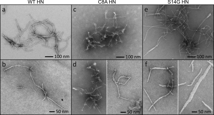
Figure 2.
Fiber formation of mutant peptides. A and B, control samples of the WT network (A) and single WT fibers (B) are imaged as before. C, the C8A mutant produced shorter, more irregularly shaped fibers and showed clear evidence of branching. Their diameters are not uniform, and the fibers form into a dense network. D, the C8A mutant cannot form into single fibers. There is always some branching or fibers are attached by globular nodes. E, the S14G mutant has similar networking properties to the WT, except there are fewer fibers running in parallel. F, single S14G fibers can appear as seen in the WT, but their diameters are larger on average ranging from 15 to 50 nm.

