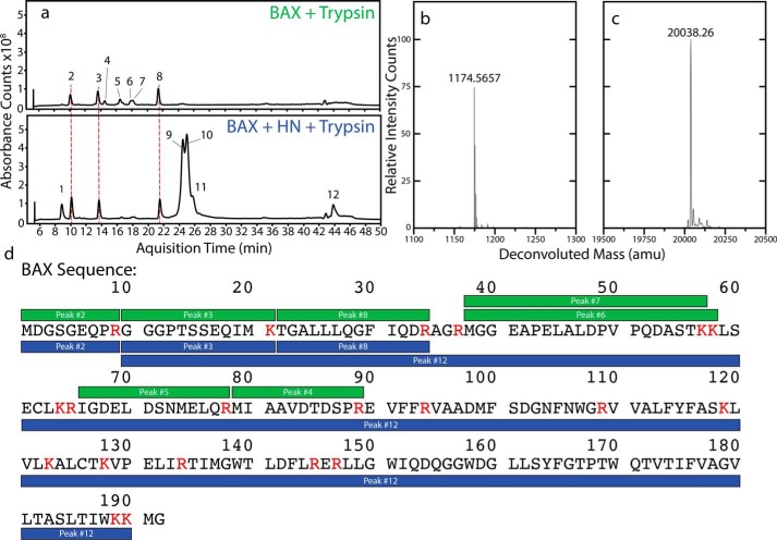
Figure 4.
Fibers protect BAX from proteolytic digestion by trypsin. A, total ion chromatograms for BAX digested with trypsin (above) and for the fiber digested under the same conditions (below). The fibers formed protect some part of BAX from digestion as evidenced by several peaks missing with the fiber samples. B, the deconvoluted MS for Peak 4 showing a typical mass intensity for a fragment that only appears in the control sample. C, the deconvoluted MS for Peak 12 showing the correct mass for a segment of BAX that is incorporated into the fiber. D, fragments identified in BAX-only control digestions (green) and digestions with fibers (blue) overlaid with the sequence of WT BAX. Residues labeled red precede trypsin cleavage sites. In the control digestions the N-terminal half of BAX can be detected in six fragments. When a fiber sample is digested, the three fragments closer to the C-terminal end of the protein are no longer observed and a new peak appears corresponding to a fragment that is nearly the full length of the protein.

