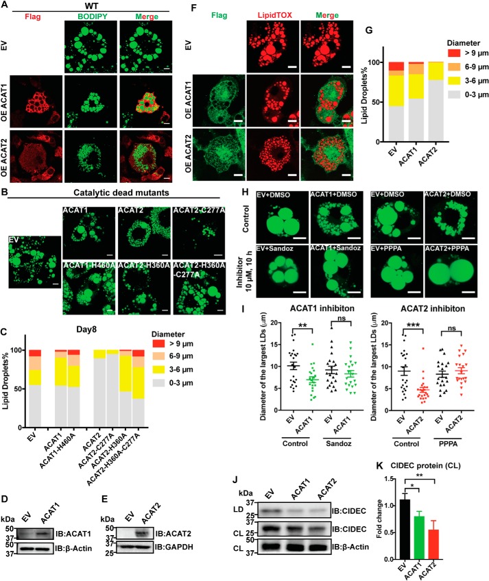
Figure 3.
ACAT1/2 overexpression impaired LD expansion in adipocytes. A, the effect of WT ACAT1/2 overexpression on LD sizes on day 8 of differentiation. Immunofluorescence was carried out with anti-FLAG primary antibody on cells expressing FLAG-tagged ACAT1/2, but not on EV control cells. LDs were stained using BODIPY. Bars, 10 μm. B, the effect of overexpressing catalytic dead mutants of ACAT1/2 on LD size on day 8 of differentiation. LDs were stained using BODIPY. Bars, 10 μm. C, quantification of LD sizes in adipocytes stably expressing ACAT1/2. LDs from ∼15 cells/cell type were used. D, protein level of ACAT1 in mature adipocytes transiently overexpressing ACAT1. E, protein level of ACAT2 in mature adipocytes transiently overexpressing ACAT2. F, the effect of transient overexpression of ACAT1/2 on LD size in mature adipocytes. Immunofluorescence was carried out with anti-FLAG primary antibody on cells expressing FLAG-tagged ACAT1/2, but not on EV control cells. LDs were stained using LipidTOX. Bars, 10 μm. G, quantification of LD sizes in mature adipocytes overexpressing ACAT1/2. LDs from ∼15 cells/cell type were used. H, the effects of ACAT1 and ACAT2 inhibitors on LD size in mature adipocytes. Bars, 10 μm. LDs were stained by BODIPY. Sandoz 58-035 (ACAT1 inhibitor) and PPPA (ACAT2 inhibitor) were dissolved in DMSO. 10 μm of inhibitor was added to respective cells for 10 h. I, quantification of diameters of 2 largest LDs in each cell type as shown in H. Two-tailed Student's t test was used (means ± S.E.; n = 20 LDs from 10 cells for each cell type). **, p < 0.01; ***, p < 0.01; ns, no significance. J, immunoblot (IB) analysis of CIDEC in cell lysates (CL) and LD fractions isolated from mature adipocytes transiently overexpressing ACAT1/2. K, quantification of CIDEC level in J. Two-tailed Student's t test was used (means ± S.D.; n = 3). *, p < 0.05; **, p < 0.01.

