Figure 5.
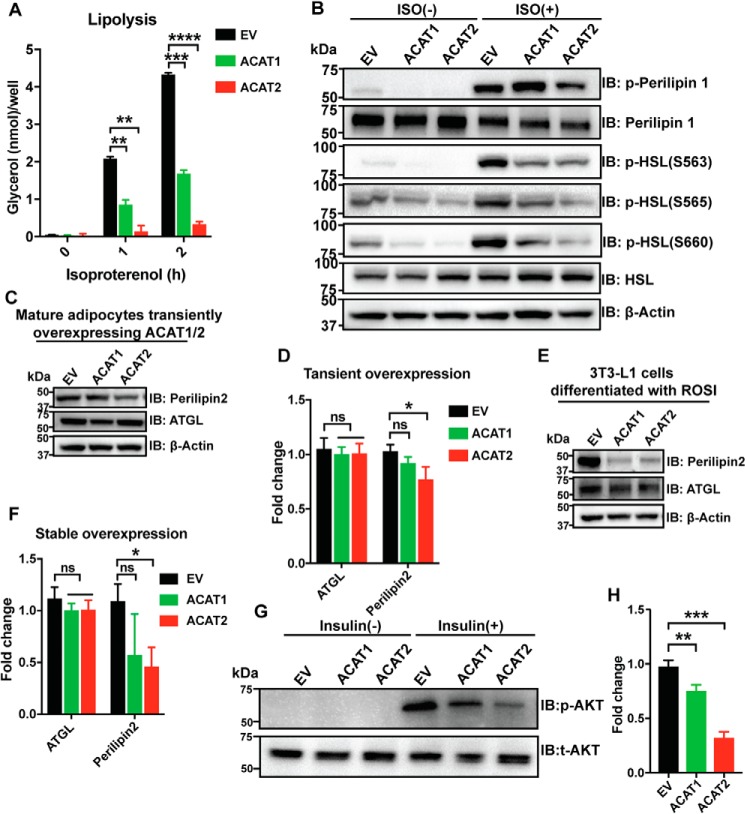
ACAT1/2 overexpression impaired metabolic functions in adipocytes. A, lipolysis assay in mature adipocytes overexpressing ACAT1/2. 100 nm isoproterenol was used. Two-tailed Student's t test was used (means ± S.D.; n = 3). **, p < 0.01; ***, p < 0.001; ****, p < 0.0001. B, immunoblot (IB) analysis of p-perilipin 1 (Ser522) and p-HSL (Ser660) in mature adipocytes transiently overexpressing ACAT1/2. ISO, isoproterenol, 10 μm, 2 h. C, immunoblot analysis of perilipin 2 and ATGL in mature adipocytes overexpressing ACAT1/2. D, quantification of perilipin2 and ATGL levels in C. Two-tailed Student's t test was used (means ± S.D.; n = 3). *, p < 0.05; ns, no significance. E, immunoblot analysis of perilipin 2 and ATGL in mature adipocytes stably overexpressing ACAT1/2. F, quantification of perilipin2 and ATGL levels in E. Two-tailed Student's t test was used (means ± S.D.; n = 3). *, p < 0.05; ns, no significance. G, immunoblot analysis of p-AKT (Ser473) in mature adipocytes stably overexpressing ACAT1/2. 10 nm insulin was added to cells for 15 min. H, quantification of p-AKT level in G. Two-tailed Student's t test was used (means ± S.D.; n = 3). **, p < 0.01; ***, p < 0.001.

