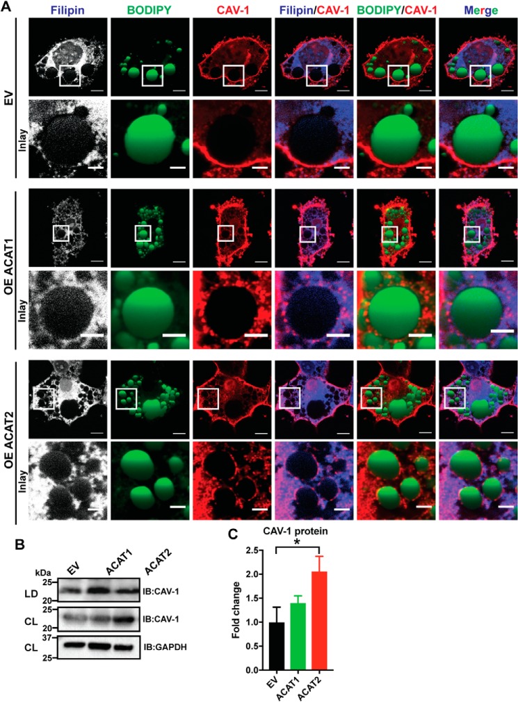
Figure 8.
ACAT1/2 overexpression promoted caveolin-1 localization to the LDs in adipocytes. A, caveolin-1 colocalized with free cholesterol on the LD surface in mature adipocytes upon ACAT1/2 overexpression. Bars, 10 μm. Inset, bars, 3 μm. CAV-1, caveolin-1. B, immunoblot analysis of caveolin-1 in LD fractions isolated from mature adipocytes transiently overexpressing ACAT1/2. CL, cell lysates. C, quantification of caveolin-1 level in B. Two-tailed Student's t test was used (means ± S.D.; n = 3). *, p < 0.05.

