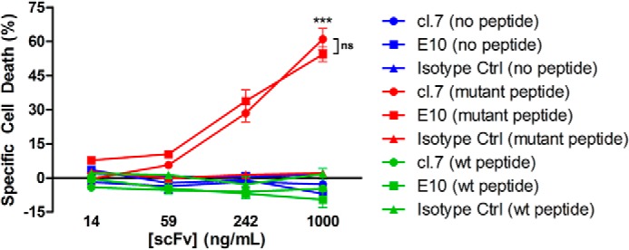
Figure 4.

ScFv-mediated, complement-dependent cell killing. T2A3 cells were peptide-pulsed with β2-microglobulin in the presence or absence of the specified peptide: no peptide (blue), the S45F mutant peptide (TTAPFLSGK) (red), or the WT peptide (TTAPSLSGK) (green). Cells were incubated with 10% rabbit serum and the cl. 7, E10, or isotype control scFvs preconjugated to anti-V5 antibody. CellTiter-Glo was used to assess the viability of cells. ***, p < 0.0001, comparing either cl.7 or E10 with isotype control at the highest antibody concentration with mutant peptide-pulsed cells; ns, not significant (p = 0.1130). Error bars, S.D.
