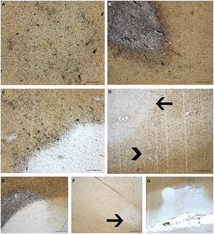
Figure 1.

Pathological characterization of MS lesions. HLA‐PLP immunohistochemistry of MS autopsy tissue showing the MS lesion characteristics. A. Reactive site with increased density of HLA+ cells with microscopically intact myelin. B. Active lesion with partial demyelination and HLA+ cells throughout the lesion. C. Mixed active/inactive (chronic active) lesion with inactive demyelinated center and a rim of HLA+ cells. D. Inactive lesion (arrow), demyelinated with sparse HLA+ cells. Remyelinated lesion (arrow head), partial myelination with sparse HLA+ cells. E. Leukocortical lesion. F. Intracortical lesion. G. Subpial cortical lesion. Scale bar represents 500 um.
