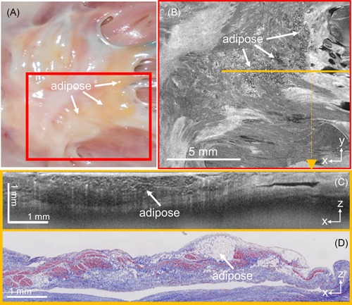
Figure 5.

Representative example of significant amounts of interstitial, subendocardial adipose at the septum primum, as imaged by optical coherence tomography (OCT). A, Camera image of region of interest. B, En face OCT image of the region depicted in the red box in A. C, OCT b‐scan corresponding to the location of the orange line in B. D, Corresponding histology to C
