Figure 1.

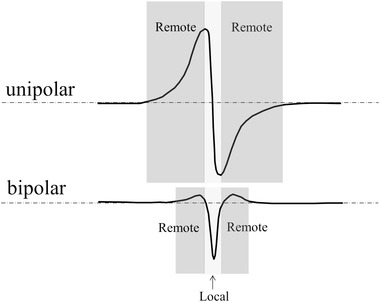
Unipolar and bipolar electrogram recorded during passage of an activation front. Parts of the extracellular electrograms reflect remote (dark gray) and local (light gray) activation. The remote part is smallest for the bipolar recording

Unipolar and bipolar electrogram recorded during passage of an activation front. Parts of the extracellular electrograms reflect remote (dark gray) and local (light gray) activation. The remote part is smallest for the bipolar recording