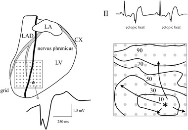
Figure 3.

Epicardial mapping in a patient with symptomatic ectopic ventricular activity (see tracing II). Anti‐arrhythmic surgery was carried out because epicardial catheter mapping revealed that the location of ectopic activity was located close to the phrenic nerve. The right lower panel shows the isochronal map of an ectopic beat derived from extracellular electrograms recorded by an 8 × 8 electrode grid (left panel). Numbers are activation times after earliest activation. The earliest activated site of the ectopic complex is indicated by an asterisk. The electrogram at that site is initially negative, as illustrated by the tracing. CX = circumflex; LA = left atrium; LAD = left anterior descending artery; RA = right atrium; LV = left ventricle; electrode diameter: 1 mm; inter‐electrode distance: 5 mm; reference electrode: surgical wound; filter setting: 0 Hz – 400 Hz
