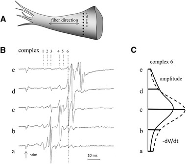
Figure 7.

Panel (A) shows a papillary muscle with a line electrode (black dots are the eight electrode terminals) positioned perpendicular to the long axis of the muscle. Unipolar electrograms recorded simultaneously at five sites (a‐e) are shown in panel (B). Numbers indicate six different complexes. Note that deflections along the dashed lines of complex 6 in the different recordings occur at the same time. Amplitude and dV/dt of these deflections are shown in panel (C). They are largest in tracing c, which suggests that the bundle that generates this deflection is located underneath electrode c. Deflections 1‐4 in tracing c have counterparts in adjacent tracings that have larger amplitude and dV/dt, thus these deflections are remote in tracing c. Deflection 5 is, however, also local because its amplitude and dV/dt are largest in tracing c. Stim., stimulus artifact; electrode diameter: 0.1 mm; inter‐electrode distance: 0.2 mm; reference electrode: at border tissue bath; filter setting: 0.01‐1 kHz
