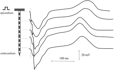
Figure 9.

Needle electrode recordings during stimulation from the epicardium. The electrode was impaled in the left ventricle of a pig heart. Note the initial negative deflection of the unipolar electrogram at the epicardial site. Activation moves to the endocardium, which is reflected by the increase in activation time. For electrograms recorded toward the endocardium, a small R‐wave arises of which the amplitude increases slightly the further the recording site is away from the epicardium
