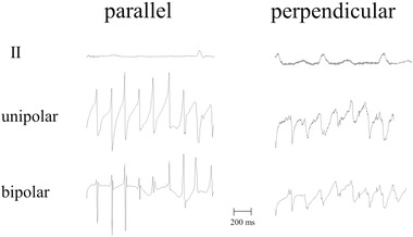
Figure 11.

Example of unipolar and bipolar recordings of a catheter with the electrode positioned parallel (left tracing) and perpendicular to the atrial wall. Recordings were made during atrial fibrillation; the bipolar tracing is the difference between the tip and the first ring electrode. If the catheter is positioned along the wall, both terminals touch the tissue and the recording is a pure bipolar one. If the catheter is, however, positioned perpendicular to the wall, only the tip touches the wall. The ring electrode records a cavity signal, which is a remote unipolar one and lower in amplitude than the tip signal. The resulting signal approaches a unipolar recording; unipolar and bipolar electrograms are similar now. Electrode size: 2 mm × 1 mm; inter‐electrode distance: 2 mm; reference electrode: vena cava inferior; filter setting: 0.5‐200 Hz
