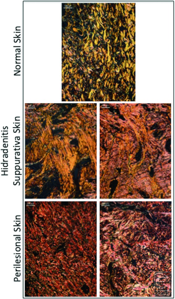
Figure 5.

Collagen-specific staining shows both type I (yellow) and III (green) collagen in normal and perilesional skin dermis, compared with predominance of type I collagen (yellow) in hidradenitis suppurativa dermis (picrosirius red stain). Scale bar = 100 µm, 10× magnification.
