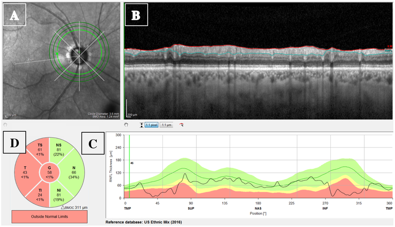
Figure 1:
The circumpapillary retinal nerve fiber layer (cpRNFL) report showing the (A) projected infrared (IR) image of the disc; (B) high-resolution circle scan with the segmented borders of the RNFL (red and blue lines); (C) cpRNFL thickness plot; and (D) Thickness of cpRNFL for temporal-inferior (TI), temporal (T), temporal-superior (TS), nasal-superior (NS), nasal (N), and nasal-inferior (NI) sectors, along with the global (G) thickness in center circle.

