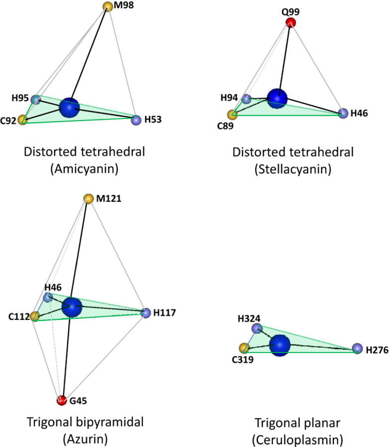
Figure 3.
Variations in the ligation geometry of type 1 copper sites. The structures were drawn using the structure coordinates from PDB files for amicyanin from Paracoccus denitrificans (PDB code 2OV0), stellacyanin from Cucumis sativus (PDB code 1JER), azurin from Pseudomonas aeruginosa (PDB code 4AZU) and ceruloplasmin from human serum (PDB code 1KCW). Only the atom providing the copper ligand is shown with the one-letter amino acid code and residue number indicated. The plane described by the two His and one Cys ligand in each is shaded light green.

