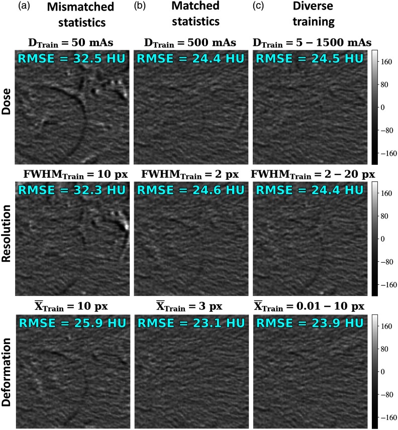
Fig. 8.
Testing on anatomical content. Difference images following registration (original images shown in Fig. 3) are shown for networks at various training conditions. RMSE of the difference in HU is shown in text for each image. Columns represent conditions of (a) mismatched training and test statistics, (b) matched statistics, and (c) diverse training. Rows examine various training conditions for dose, resolution, and deformation magnitude.

