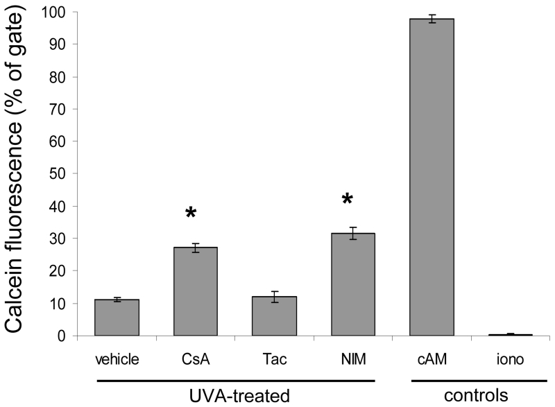
Fig. 4.
Mitochondrial Permeability Transition Pore activity is diminished in UVA-irradiated cells treated with CypD-binding drugs. The MPTP was monitored by quantifying the fluorescence of calcein in the mitochondria of HaCaT cells by flow cytometry. Cells were treated with vehicle, CsA, Tac, or NIM at 125 nM and irradiated with a single dose of 12 J/cm2 of UVA. The cells were then loaded with calcein AM (cAM) and CoCl2 (cytosolic calcein quencher) to determine the calcein fluorescence in the mitochondria. Control cells were loaded with cAM alone cAM, CoCl2, and ionomycin (iono) which triggers pore opening and loss of mitochondrial calcein fluorescence. Data represent means ± s.e.m. n=3. Asterisks indicate significant differences compared to vehicle treated cells, *P < 0.05.

