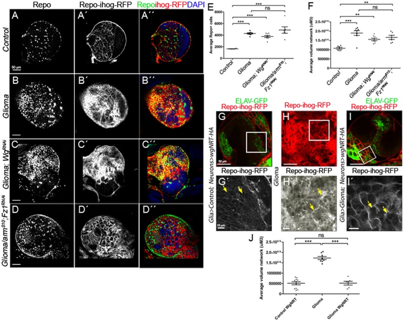
Fig 4. Wg expression in glioma cells is dispensable for tumor progression because glioma depletes Wg from neuronal membrane.
(A–F) Larval brain sections with glial membrane projections labeled in gray (red in the merge) and glial cell nuclei stained with Repo (gray, green in the merge). (C) wg knockdown in glioma cells (wg-RNAi) or armS10; Fz1-RNAi (D) does not prevent glioma cell number increase nor glioma TM volume expansion quantified in (E–F). (G–Gʹ) Control brains express 1 copy of WgNRT-HA instead of endogenous secretable Wg in neurons (green, ELAV-GFP). (H–Hʹ) Glioma samples that do not express WgNRT in neurons show an overgrown TM network. (panel I in red, magnification in panel Iʹ in gray) Glioma brains expressing membrane-anchored Wg (WgNRT-HA) in neurons show that the glial network volume size is restored to control volume in these animals. Quantification of the TM network volume is shown in panel J. Yellow arrows show glial network. Error bars show SD; ***P < 0.0001; **P < 0.001; and ns for nonsignificant. The data underlying this figure can be found in S1 Data. Genotypes: (A) w;; repo-Gal4, ihog-RFP/UAS-lacZ, (B) UAS-dEGFRλ, UAS-dp110CAAX;; repo-Gal4, UAS-ihog-RFP, (C) UAS-dEGFRλ, UAS-dp110CAAX; repo-Gal4/UAS-wg-RNAi, (D) UAS-armS10/UAS-dEGFRλ, UAS-dp110CAAX; UAS-Fz1-RNAi; repo-Gal4, UAS-ihog-RFP.UAS-ihog-RFP, (G) w; >wg>wgNRT-HA, PaxRFP/ elav-lexA, lexAop-CD8-GFP; repo-Gal4, UAS-ihog-RFP/lexAop-flp, (H) UAS-dEGFRλ, UAS-dp110CAAX; >wg>wgNRT-HA, PaxRFP; repo-Gal4, UAS-ihog-RFP/lexAop-flp, (I) UAS-dEGFRλ, UAS-dp110CAAX; >wg>wgNRT-HA, PaxRFP/ elav-lexA, lexAop-CD8-GFP; repo-Gal4, UAS-ihog-RFP/lexAop-flp. ELAV-GFP, embryonic lethal abnormal vision green fluorescent protein; Fz1, Frizzled1; TM, tumor microtube; Wg, wingless.

