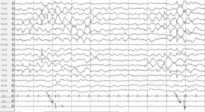
Figure 3.

Electroencephalogram of the patient at 2.5 months in awake state showing clusters of bilateral high‐amplitude central spikes followed by high‐amplitude slow waves and erratic focal right myoclonus (arrows). Time constant: 10 sec. Amplitude: 100 µV/cm. High band filter: 0.3 Hz. Low band filter: 70 Hz.
