Figure 7.

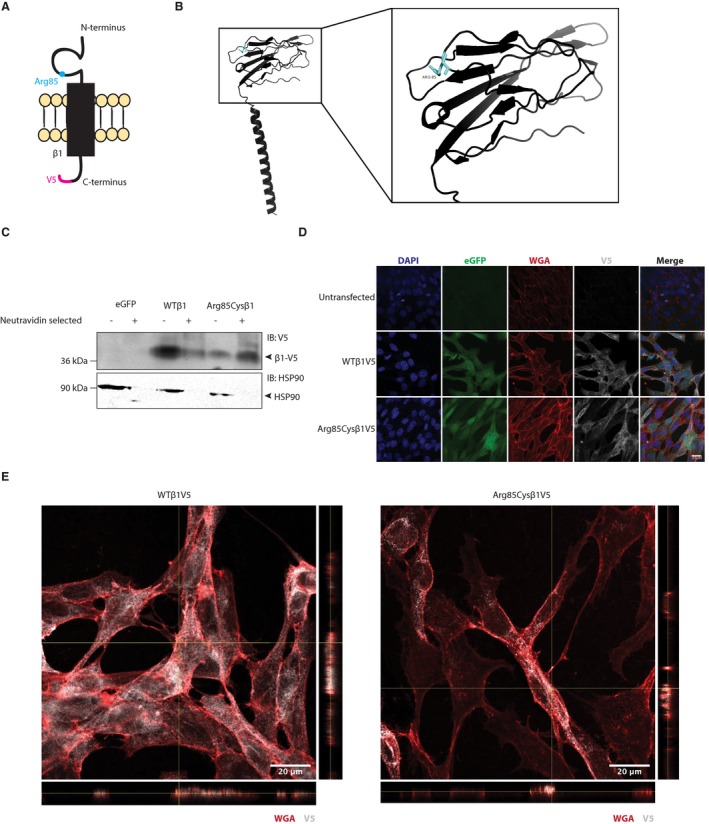
β1‐p.Arg85Cys localizes to the plasma membrane. (A) Cartoon diagram of β1‐p.Arg85Cys. (B) Crystal structure of WTβ1 (PDB: 6AGF).23 The residue, Arg85, is shown in cyan. Right: 20 angstrom area showing detail of the Ig domain. (C) Cell surface biotinylation shows that β1‐p.Arg85Cys can localize to the plasma membrane, similar to WT (representative of four independent experiments). (D) β1 WT and β1‐p.Arg85Cys colocalize with the plasma membrane marker, WGA (representative of five independent experiments). (E) Orthogonal views of a single z‐stack (YZ plane to right, XZ plane below) from immunofluorescence microscopy indicating colocalization between V5 and WGA signals.
