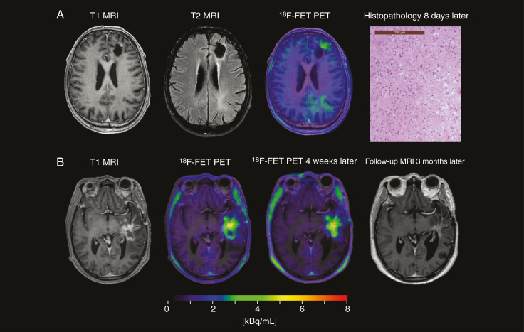
Fig. 3.
False-negative and false-positive18F-FET PET scans. (A) A 62-year-old patient with a left-sided frontal glioblastoma, treated with gross tumor resection and Stupp. Eleven months after adjuvant TMZ, MRI showed a nonmeasurable solitary contrast enhancement at the anterior part of the surgical cavity, identified by 18F-FET PET with TBRmax of 1.9 and BTV of 0.5 cm3. A suspicion of disease recurrence was raised due to the nodular configuration. Subsequent histopathology showed recurrent glioblastoma (magnification x200). (B) A 48-year-old patient with a left-sided temporal glioblastoma, treated with subtotal tumor resection and Stupp, and re-resection 5 months later with histopathological findings of posttreatment changes. Nine months after radiotherapy, MRI demonstrated progressive contrast-enhancing lesion near the surgical cavity. Two consecutive 18F-FET PET with a 4-week window showed increased uptakes, with TBRmax of 3.1 and 3.5, respectively. Three months later, a spontaneous regression was observed.

