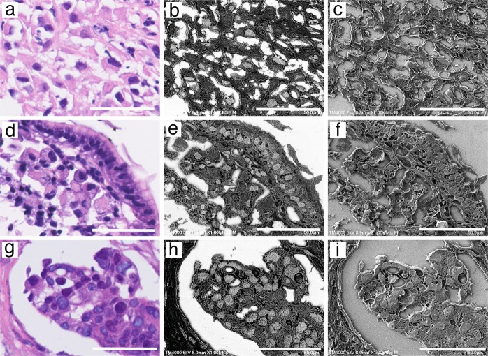
Fig. 4.
Identification of the nucleus in paraffin sections with gold (III) chloride using the NanoSuit method. Microscopic examination of: a–c malignant mesothelioma sample, d–f gastric carcinoma sample (signet ring cell carcinoma), and g–i breast cancer sample. The panels represent: hematoxylin and eosin (H&E) staining (a, d, g), Low-vacuum scanning electron microscopy (Lv-SEM) image of a sample stained using 1% gold (III) chloride, taken in BSE mode (b, e, h), and mixed Lv-SEM image of a sample stained with 1% gold (III) chloride (c, f, i). The scale bars represent 50 μm

