Figure 1.
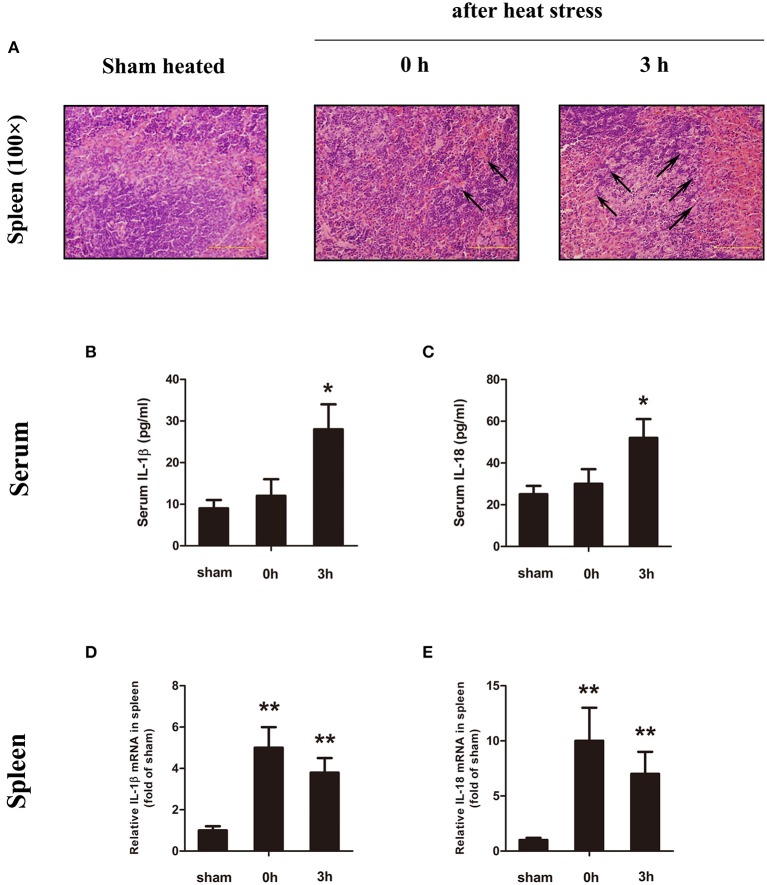
Heatstroke (HS) induced spleen injury and inflammatory cytokine upregulation. (A) Representative pathological images of the spleen from sham heated mice (left) and HS mice (0 h, middle; 3 h, right) stained with H&E at a magnification of ×100. At the time of HS (0 h), the body temperature was high (Tc = 43°C), the spleen sinus expanded, and the red pulp region showed obvious congestion (black arrow), but no obvious cell damage and changes were observed. Three hours after the occurrence of HS (low-body-temperature period, Tc = 29°C), the splenic sinus congestion was severe (black arrow), and lymphocyte disintegration led to increased cell debris in the red pulp and phagocytosis by phagocytic cells. In each group, n = 6. H&E, hematoxylin and eosin. (B–E) Interleukin (IL)-1β and IL-18 protein expression in serum (B,C) measured by enzyme-linked immunosorbent assay (ELISA) at the time point of HS onset (0 h) and 3 h after onset. Meanwhile, the spleen (D,E) was measured by real-time quantitative PCR at the time point of HS onset (0 h) and 3 h after onset. Means and standard deviations are shown, n = 6 per group, *p < 0.05 compared with the sham heated group, **p < 0.001 compared with the sham heated group.

