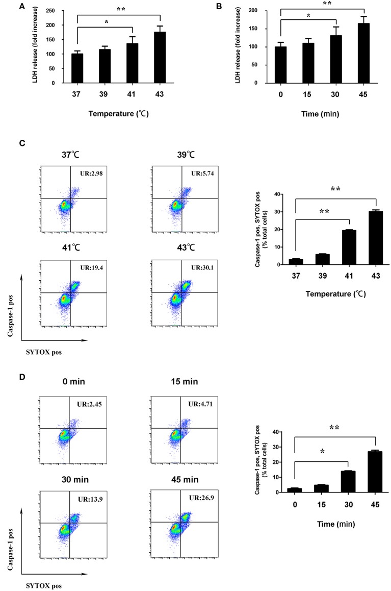
Figure 4.
Heat stress destroyed cell membrane integrity of splenic lymphocytes in vitro. (A) Cells were incubated at different temperatures (37, 39, 41, and 43°C) for 45 min. Levels of lactate dehydrogenase (LDH) release are shown in the bar graph. (B) Cells were subjected to heat stress (43°C) for different times (0, 15, 30, and 45 min). Levels of LDH release are shown in the bar graph. (C) Cells were incubated at 37°C (control) and at different temperatures (39, 41, and 43°C) for 45 min; effects of different temperatures on the caspase-1 (FLICA 660-YVAD-FMK staining, vertical) and SYTOX Green (horizontal) double-positive cells measured by flow cytometry. The ratio of caspase-1 and SYTOX Green double-positive cells is shown in the bar graph. (D) Cells were subjected to heat stress (43°C) for different times (0, 15, 30, and 45 min). Effects of different durations on caspase-1 (FLICA 660-YVAD-FMK staining, vertical) and SYTOX Green (horizontal) double-positive cells under high temperature measured by flow cytometry. The ratio of caspase-1 and SYTOX Green double-positive cells is shown in the bar graph. Values are mean ± SD for six independent experiments. *p < 0.05, **p < 0.01 compared with the control group (37°C) or sham heated group.

