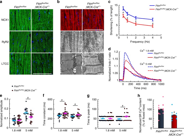
Fig. 2. Impaired myocyte function in 10-month-old MCK-Fktn-cKO (Fktnflox/flox; MCK-Cre+/−) mice.
a Representative immunostaining for NCX1, RyR2, and LTCC (DAPI counterstain, blue) in hearts of 10-month-old mice. Scale bar, 100 μm. b Representative immunofluorescence (top: phalloidin, red; DAPI, blue (scale bar, 100 μm)) and electron microscopy (scale bar, 5 μm (middle) and 1 μm (lower)) images of myofilaments. c Frequency-dependent shortening of cardiomyocytes (n = 16 and 20 cells measured from 4 and 5 hearts, respectively). #P < 0.05 vs. floxed mice based on Student’s t-tests. (d) Indo-1 fluorescence in single cardiomyocytes stimulated at 1 Hz (1.8 mM Ca2+, n = 20 and 37 cells from 3 and 4 hearts, respectively; 5 mM Ca2+, n = 20 cells from 3 hearts per group). Normalized peak amplitudes (e), decay time constants (obtained by fitting to the decline phase) (f), and times to peak (g) of Ca2+ transients. h Estimation of sarcoplasmic reticulum (SR) Ca2+ content (n = 15 and 10 cells from 3 and 2 hearts, respectively). #P < 0.05 between indicated groups based on Student’s t-tests.

