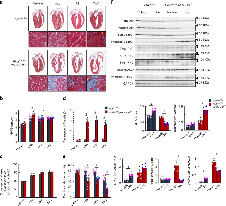
Fig. 3. Hypertrophic responses of 10-week-old MCK-Fktn-cKO (Fktnflox/flox; MCK-Cre+/−) hearts.
a Cardiac morphology and histology following isoproterenol (Iso) or phenylephrine (PE) administration or thoracic aortic constriction (TAC) for 2 weeks in control (floxed) and MCK-Fktn-cKO mice. Scale bar, 5 mm (top) and 50 μm (bottom). b Heart weight-to-body weight ratios (HW/BW) (n = 9 mice per group). Cross-sectional areas (vehicle, n = 252 cells; +Iso, n = 283 cells; +PE, n = 249 cells; TAC, n = 264 cells from 3 control (floxed) mice. Vehicle, n = 315 cells; +Iso, n = 250 cells; +PE, n = 229 cells; TAC, n = 274 cells from 3 MCK-Fktn-cKO mice) (c) and fibrosis percentages (n = 3 mice per group) (d) from paraffin sections of left ventricles. e Fractional shortening in hearts of treated and untreated control (n = 12 mice per group). *P < 0.05 vs. vehicle-treated floxed mice; #P < 0.05 between indicated groups based on Tukey–Kramer tests. f Representative immunoblots for total and phosphorylated Akt, CamKII, PKD, and HDAC9 levels in control and MCK-Fktn-cKO mice after Iso (or vehicle) treatment. GAPDH was used for a loading control (n = 6 per group). *P < 0.05 vs. vehicle-treated floxed mice; #P < 0.05 between indicated groups based on Tukey–Kramer tests.

