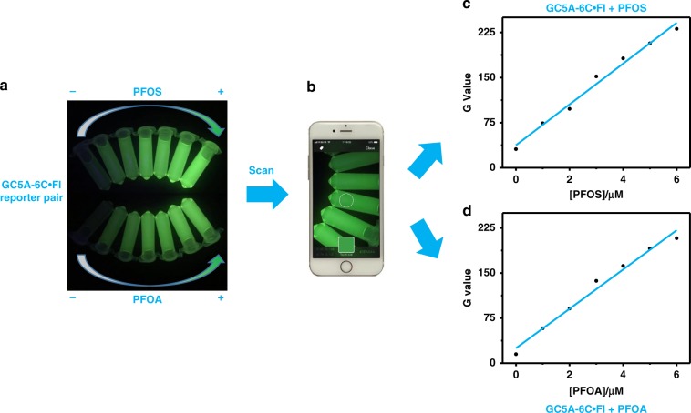
Fig. 6. Real-time/on-site scanometric monitoring of PFOS and PFOA.
a The images of the GC5A-6C•Fl reporter pair (8.0/10.0 μM) with various concentrations of PFOS and PFOA (up to 6.0 μM) taken by an iPhone 7. b The images recorded by iPhone 7 with a color-scanning app. Plots of G values against PFOS (c) and PFOA (d) concentrations in HEPES buffer at 25 °C. G values are green color intensities directly scanned from the color scanning app.

