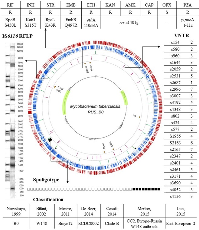
Figure 1.
Circular map and genetic features of M. tuberculosis RUS_B0. Two outer circles show the coding sequences on plus (red) and minus (blue) strands; the third ring depicts CRISPR locus (black) and IS6110 integration sites (green) (Beijing B0/W148 specific sites indicated by asterisk); the fourth ring shows the positions of the VNTR; the fifth ring shows drug resistance genes; and the sixth ring shows the scale in the genome with the inverted regions with respect to the H37Rv genome.

