Figure 1.
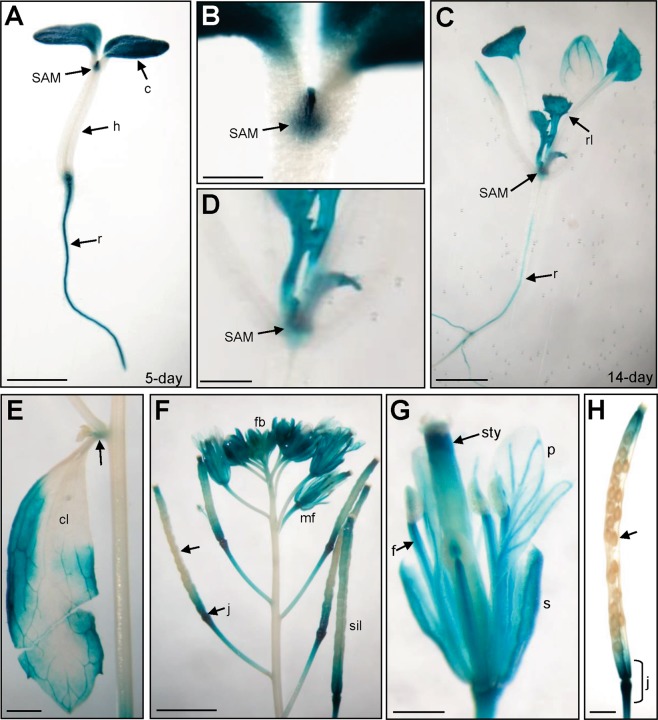
GUS staining patterns in GSF::GUS transgenic Arabidopsis. (A) GUS activity was specifically detected in the cotyledon (c), shoot apex meristem (SAM) and roots (r) of a 5-day-old seedling. GUS was absent in the hypocotyl (h). Bar = 3 mm. (B) Close-up of the shoot apex meristem (SAM) region from (A). Bar = 0.5 mm. (C) GUS activity was specifically detected in the shoot apex meristem (SAM), rosette leaves (rl) and roots (r) of 14-day-old seedlings. Bar = 3 mm. (D) Close-up of the shoot apex meristem (SAM) region from (C). Bar = 1 mm. (E) GUS activity was detected in the cauline leaf (cl) and the node of inflorescence (arrow) in the inflorescence of a mature plant. Bar = 1 mm. (F) In a mature inflorescence, GUS activity was detected in the flower organs in both flower buds (fb) and mature flowers (mf). In siliques, GUS was strongly detected in the junction region (j) of the silique and pedicel and was absent from the developing seeds (arrow). Bar = 25 mm. (G) In a mature flower from (F), GUS activity was detected in the sepal (s), stamen filaments (f) and style of the carpel (sty) and weakly detected in the petals (p). Bar = 5 mm. (H) In a mature silique from (F), GUS was strongly detected in the junction region of the silique and pedicel (j) and was absent in the developing seeds (arrow). Bar = 0.5 mm.

