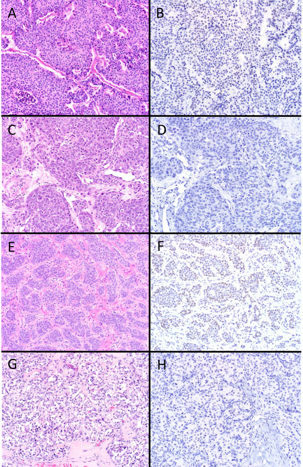
Figure 3.
SATB2 “Rarely Positive” Well-Differentiated Neuroendocrine Neoplasms. Around 10% of bronchopulmonary, thyroid and jejunoileal NETs and paragangliomas were SATB2-positive, with H-scores typically in the single digits to few dozen. Several of the strongest “aberrant” expressors are depicted here. Atypical carcinoid tumors of lung origin (A; note focus of punctate necrosis at the lower left) were more likely to be SATB2-positive (B) than typical carcinoid tumor. A medullary thyroid carcinoma (C) demonstrating rare cells staining (D). Ileal NET (E) demonstrating moderately strong staining (F); of note, this tumor was encountered clinically subsequent to the completion of the study and represents the strongest SATB2-positivity I have seen in a NET outside of the appendix and rectum. Paraganglioma (G) displaying rare cells staining (H) (original magnification of each image 200x).

