
Figure 2.
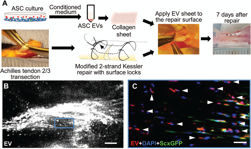
Application and localization of ASC EVs in a mouse model of Achilles tendon injury and repair. (A) A schematic illustration of the process of preparation and in vivo application of ASC EVs in a mouse model of Achilles tendon partial transection and repair. (B) A representative whole-mount fluorescence image showing the injury site of a mouse Achilles tendon 7 days after partial transection and application of ASC EVs. Scale bar, 100 μm. (C) A fluorescence image showing the sagittal section of the tendon shown in B at the boxed region. The residing tenocytes expressed ScxGFP. All cells were counterstained with DAPI. The arrow heads point to the EV positive signals at the DAPI positive and ScxGFP negative cells. Scale bar, 50 μm.