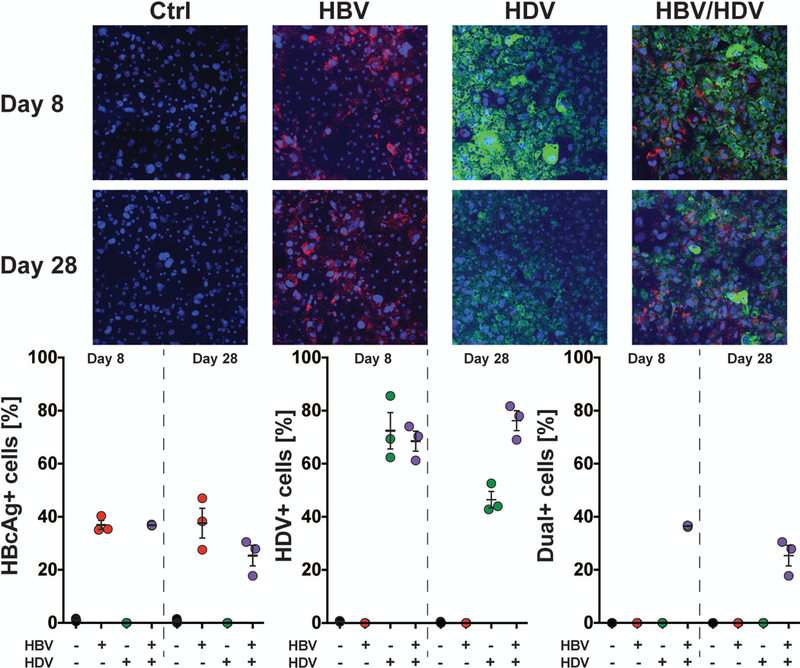
Fig. 2. Quantification of infection in HBV, HDV, and co-infected SACC-PHHs by HDV vPLAYR/TSA and anti-HBcAg immunofluorescence staining.

SACC-PHHs were either infected with HBV (MOI=4,000), HDV (MOI=1,000), or both HBV/HDV (HBV MOI=4,000, HDV MOI=1,000). (A) At 8 (top) and 28 (bottom) days post infection, control and infected SACC-PHHs were fixed and stained for HDV genomic RNA (green) by a vPLAYR/TSA procedure and for HBcAg (red) by an anti-HBcAg antibody as well as with DAPI (blue) for nuclear DNA. Quantification of three different images for each experimental condition were performed (approximately ~800 cells total per condition) for (B) HBcAg positive cells; (C) HDV genomic RNA positive cells; and (D) dual-positive cells.
