FIG 1.
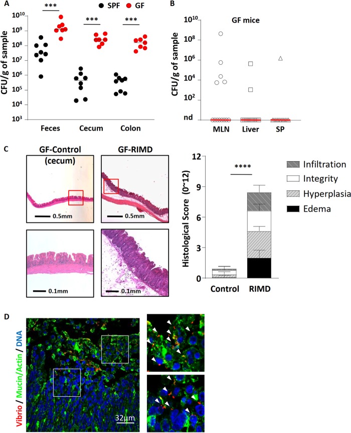
V. parahaemolyticus RIMD2210633 heavily colonizes the intestines and causes cecitis in germfree (GF) mice. V. parahaemolyticus was orally gavaged into germfree C57BL/6 mice that were euthanized at 21 h p.i. for analysis. (A) Colonization (CFU) of V. parahaemolyticus was measured from feces and cecal and colonic tissue and compared to streptomycin-pretreated SPF mice that were infected with V. parahaemolyticus. (B) V. parahaemolyticus RIMD rarely translocated into systemic tissues: mesenteric lymph nodes (MLN), liver, and spleen (SP). nd, not detected. (C) Representative H&E staining images and histologic scores of infected and uninfected ceca of germfree mice. (D) Representative immunostaining images of cecum show V. parahaemolyticus RIMD (red; noted by white arrowheads) invasion into cecal tissues. Mucin and actin are shown in green, and DNA is shown in blue. In the graphs, bars show the median (A and B) or the mean ± SEM (C), and each symbol represents an individual mouse from two independent experiments. ***, P < 0.001, and ***, P < 0.0001, by Mann-Whitney test (A and B) or Student’s t test (C).

