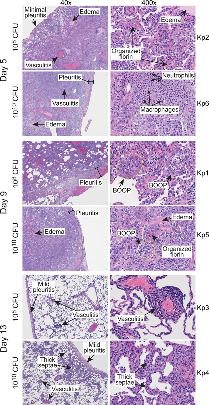
FIG 2.

Histopathology of lungs infected with K. pneumoniae. Images represent sections of lung from cynomolgus macaques infected with 108 or 1010 CFU of K. pneumoniae strain ST258 as indicated. Tissue sections were collected during necropsy on the indicated day postinfection and stained with hematoxylin-eosin. The original magnification is 40× (left) or 400× (right). BOOP, bronchiolitis obliterans organizing pneumonia. Kp1 to Kp6 indicate numbers of individual animals.
