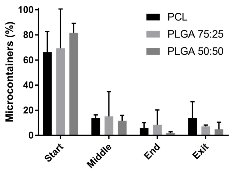
Figure 3.
Percentage of microcontainers located in the start, middle, end, and exit of the small intestine of a pig after an ex vivo perfusion study. Comparison of poly-ɛ-caprolactone (PCL) (black) microcontainers, PLGA 75:25 (dark grey), and PLGA 50:50 (light grey). Data is presented as mean ± SD with n = 3–4.

