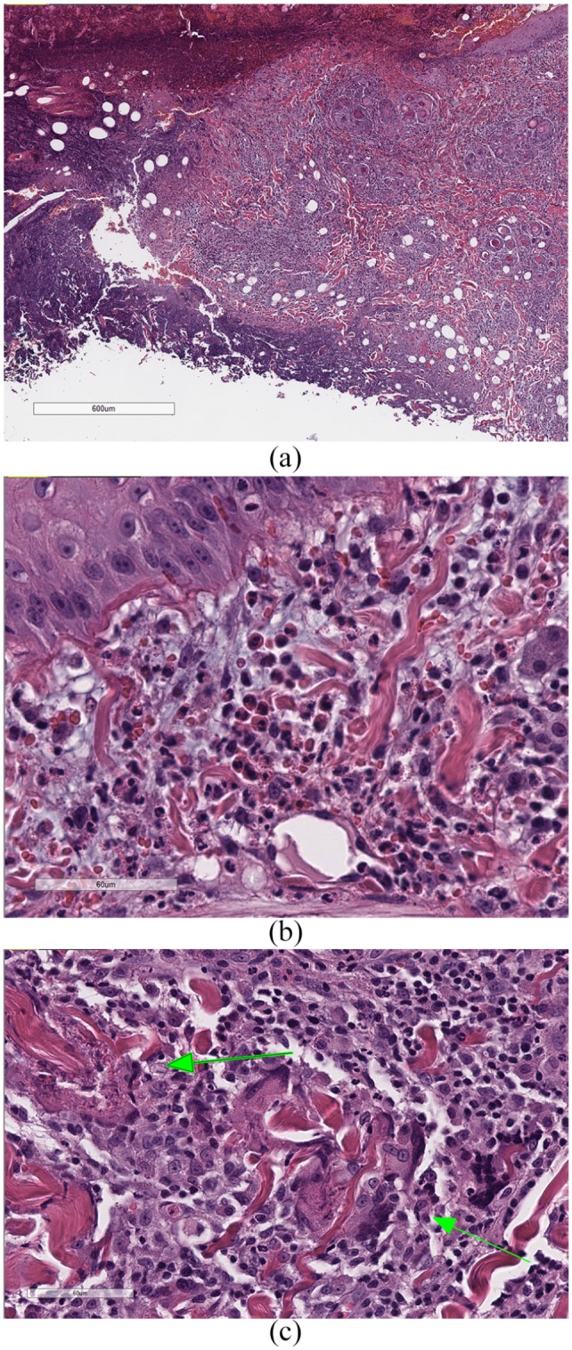
Figure 2.

(a) Ulcerative dermatitis with necrosis of the underlying dermis, and heavy dermal infiltrates of eosinophils; (b) dermal infiltrates of eosinophils, inflammatory mast cells and small lymphocytes, with mild-to-moderate edema and mucinosis; and (c) ‘flame figures’ (arrows), consisting of collagen bundles surrounded by degranulated eosinophils, which, in turn, are surrounded by macrophages and multinucleated giant cells. The bars in (a), (b) and (c) are 600 μm, 60 μm and 60 μm, respectively
