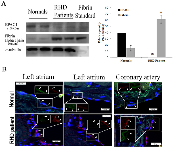
Fig. 2.
Reduced expression of EPAC1 and increased expression of fibrin in endocardial tissues underneath atrial mural thrombi in humans. Representative WB (A) shows markedly reduced expression of EPAC1 and increased expression of fibrin in endocardial areas (including cardiac endothelium) underneath atrial mural thrombi from patients with rheumatic heart disease (RHD) mitral stenosis with chronic atrial fibrillation compared to normal donor heart cases. Densitometry was used to quantify the relative intensity of EPAC1- and fibrin-specific immunoblots from patients (n = 4) and normal cases (n = 4) normalized by α-tubulin-specific controls. EPAC1 levels are presented as percentages of the indicated loading controls (*P < 0.01, compared to normal control). Representative dual-target IF staining localizes EPAC1 (green) and fibrin(ogen) (red) in areas (arrowheads) of right atrial endocardium and intima layer of coronary blood vessel walls (B). Inserts depict split signals of EPAC1 (green) and fibrin(ogen) (red) from the same area. Standard: human fibrin protein (Sigma-Aldrich). Scale bars indicate 20 μm.

