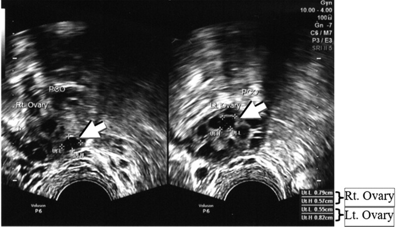
FIG. 1.
Follicle growth in polycystic ovary syndrome (arrows). Follicle sizes were measured for Ut-L (follicle length), diameter 1; and Ut-H (follicle height), diameter 2. This transvaginal ultrasound was taken on day 2 of menstruation. A diameter of 0.68 cm in the right ovary was seen clearly (0.79 + 0.57)/2 = 0.68, and there was a diameter of 0.69 cm in the left ovary (0.55 + 0.82)/2 = 0.69.

