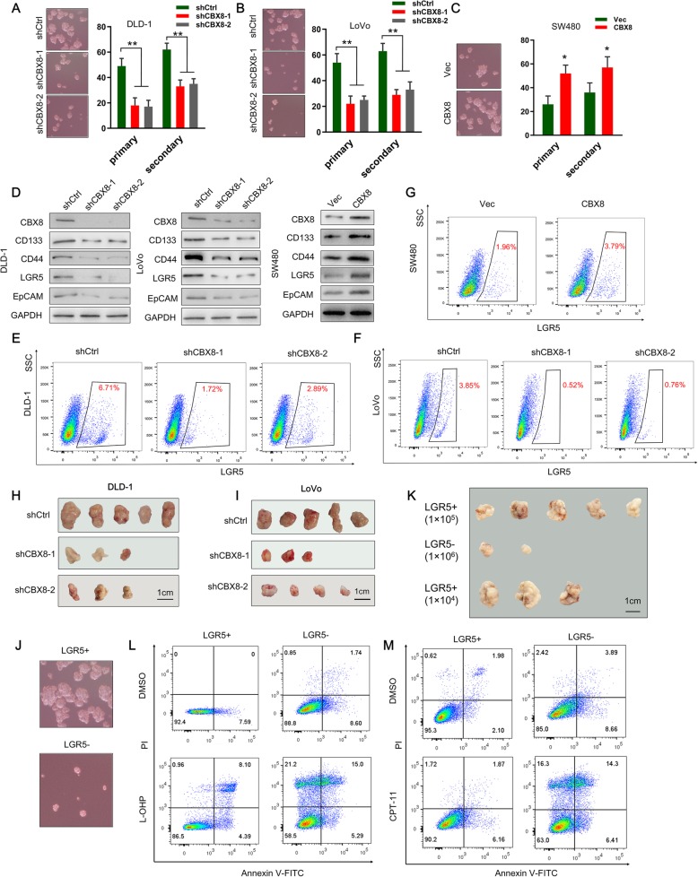
Fig. 1.
CBX8 maintains the stemness of CC cells. a-c, Representative images of sphere formation induced by the transfection of shCBX8 into DLD-1 and LoVo cells or the transfection of a CBX8 overexpression plasmid into SW480 cells. The surviving colonies were measured for the number of tumorspheres. d, The expression levels of CSC markers, including CD133, CD44, LGR5 and EpCAM, were examined in shCBX8-transfected CC cells and CBX8 overexpression plasmid-transfected CC cells by Western blotting. e-g, Flow cytometry was used to assess the percentage of LGR5high cells in CC cells with CBX8 depletion or overexpression. h-i, Tumor formation in nude mice injected with shCBX8-transfected DLD-1 and LoVo cells (5 × 10⁴ cells per mouse). The incidence of tumor formation was monitored for 40 days. j, Sphere formation of sorted LGR5+ and LGR5- DLD-1 cells. Only the top 2% most brightly stained cells or the bottom 2% most dimly stained cells were selected as LGR5+ or LGR5- populations, respectively. k, Xenograft tumors derived from serial subcutaneous injections of sorted LGR5+ and LGR5- DLD-1 cells. l-m, Sorted LGR5+ cells and LGR5- cells were treated with L-OHP and CPT-11. Apoptotic rate was detected by double-stained for Annexin V and PI and analyzed by flow cytometry. Data are shown as the mean ± SD of three replicates (*, P < 0.05; **, P < 0.01)

