Fig. 1.
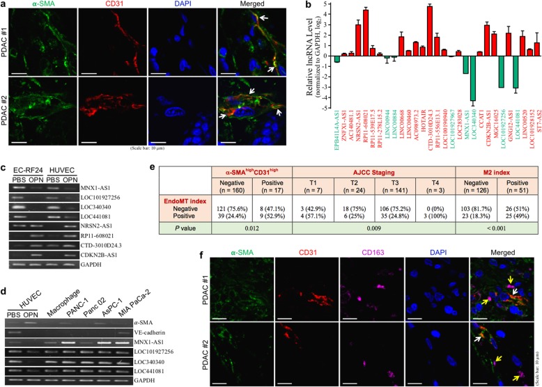
EndoMT occurs significantly with M2-macrophage infiltration in PDAC tissues. a IHF of α-SMA and CD31 from the tumor tissues of PDAC patients. Nuclei were stained with DAPI. EndoMT-derived cells exhibiting α-SMA+ and CD31+ are indicated by arrows. b Changes of the lncRNA levels in EndoMT cells derived from HUVECs. Among the 29 lncRNAs analyzed by qPCR, 21 of them were upregulated and only 8 were downregulated. c Downregulation of MNX1-AS1, LOC101927256, LOC340340, and LOC441081 and upregulation of RP11-608021, CTD-3010D24.3, and CDKN2B-AS1 in EndoMT cells derived from OPN-treated HUVECs and human immortalized endothelial cell line EC-RF24 cells. d Low expression of MNX1-AS1, LOC101927256, LOC340340, and LOC441081 in HUVEC-derived EndoMT cells compared with PDAC cells and macrophages. e Correlations of the EndoMT level of PDAC tissues with T4 staging and M2-macrophage infiltration. The data of 177 PDAC patients in TCGA database were analyzed to reveal the clinical relevance of the EndoMT occurrence. Using average expression values as cut-off points, we proposed a combination of low expressions (< averages) of LOC340340, LOC101927256, and MNX1-AS1 as the EndoMT index, and observed that PDAC tissues with positive EndoMT index were significantly correlated with T4 staging and positive for M2-macrophage index (CD163high and CD204high). The data of AJCC staging were available from 175 patients. f IHF of α-SMA, CD31, and CD163 showing EndoMT-derived cells (indicated by white arrows) and neighboring M2-type macrophages (indicated by yellow arrows) in the tumor tissues of PDAC patients. Nuclei were stained with DAPI

