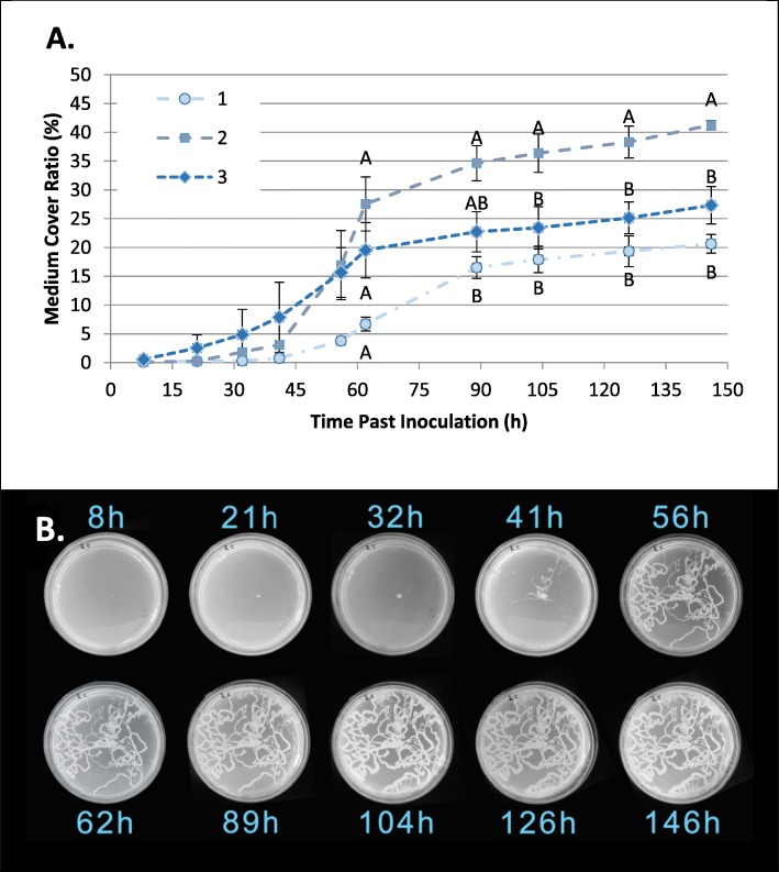
Fig. 3.
Larvae-mediated dispersal of bacteria. a Bacterial growth, measured as a function of time (as % of total surface) following the placement of one, two or three medfly eggs onto a Petri dish containing solid LB is presented as % of total surface area. Differences between groups were established separately for each time point. Different letters denote significant differences between groups for each time point (Tukey’s HSD P < 0.05). b Time-lapse photographs of a single plate containing two larvae. The spread of bacteria is clearly visible by trails of developing colonies depicting the movements of advancing larvae

