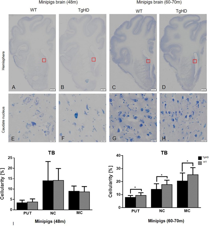
Fig. 7.
Toluidine Blue histochemical staining and quantification of cellularity. Hemispheres (A-D); caudate nucleus (E-H). (I) Quantification of cellularity in striatum and motor cortex of minipig brain sections of both 48- and 60- to 70-month-old animals using image analysis methods. Significantly decreased cellularity was detected in the putamen (PUT), caudate nucleus (NC) and motor cortex (MC) of TgHD 66-month-old animals. *P≤0.05. TB, Toluidine Blue. Scale bars: hemispheres, 2 mm; enlargements of brain structures, 50 µm.

