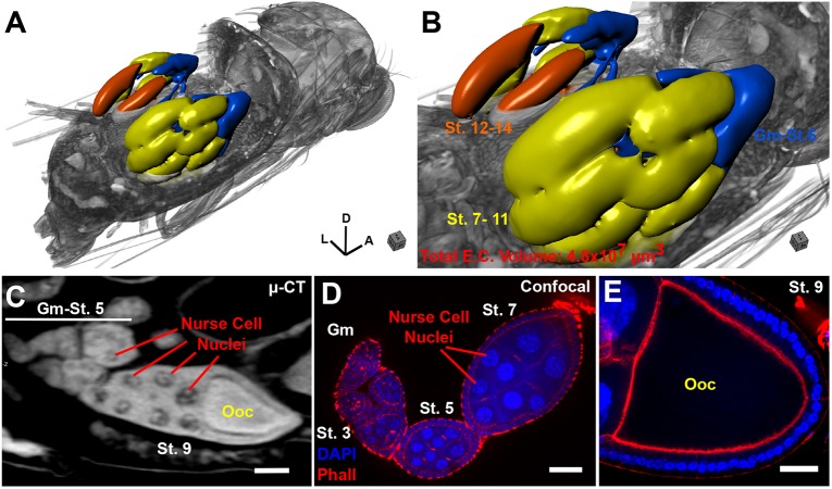
Fig. 5.
µ-CT of adult Drosophila melanogaster highlighting the female reproductive system. (A) 3D view of a female fruit fly, with the abdomen digitally removed to reveal the underlying structure of the ovarioles within the ovary. Body axes are denoted: A, anterior; D, dorsal; L, left. (B) Close-up of A; ovarioles are rendered as a surface and colored according to oogenesis stage [blue, germarium (Gm)-Stage (St.) 6); yellow, Stage 7-11; orange, Stage 12-14]. Total egg chamber volume is shown. (C) 2D image of an egg chamber imaged by µ-CT. Stages are denoted along with position of the oocyte (Ooc) and nurse cell nuclei (note visualization of polytene chromosomes). (D) Confocal image of an ovariole stained with DAPI and phalloidin; stages are indicated. (E) Confocal image of an oocyte from a stage 9 egg. Scale bars: 50 µm (C-E). Stained with 0.1 N iodine and scanned in slow mode at an image pixel size of 1.25 µm.

