Fig. 3.
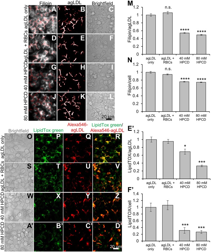
Cholesterol-balanced HPCD reduces FC uptake from untethered agLDL and foam cell formation. HPCD was pre-incubated with RBCs to cholesterol balance HPCD. J774 macrophages were incubated with Alexa546–agLDL for 1 h and then subsequently for 3 h with (A–C) no further treatment, (D–F) RBCs alone, (G–I) RBCs and 40 mM HPCD or (J–L) RBCs and 80 mM HPCD. The cells were fixed and FC stained with filipin and analyzed by confocal microscopy. Confocal images were used to quantify the average filipin intensity in (M) agLDL and in (N) cells, and (M) agLDL size for at least 10 fields containing >100 cells relative to that in control cells (agLGL only, set at 1). (O–F′) HPCD was pre-incubated with RBCs to cholesterol balance HPCD. J774 macrophages were incubated with Alexa546–agLDL for 1 h and then subsequently for 3 h with (O–R) no further treatment, (S–V) RBCs alone, (W–Z) RBCs and 40 mM HPCD or (A′–D′) RBCs and 80 mM HPCD. The cells were fixed and cholesteryl esters stained with LipidTox Green and analyzed by confocal microscopy. Confocal images were used to quantify LipidTox Green intensity in (E′) agLDL and in (F′) cells for at least 10 fields containing >100 cells relative to that in control cells (agLGL only, set at 1). Data were compiled from at least three independent experiments. Error bars show s.e.m. *P≤0.05, ***P≤0.001 ****P≤0.0001; n.s. not statistically significant.

