Fig. 1.
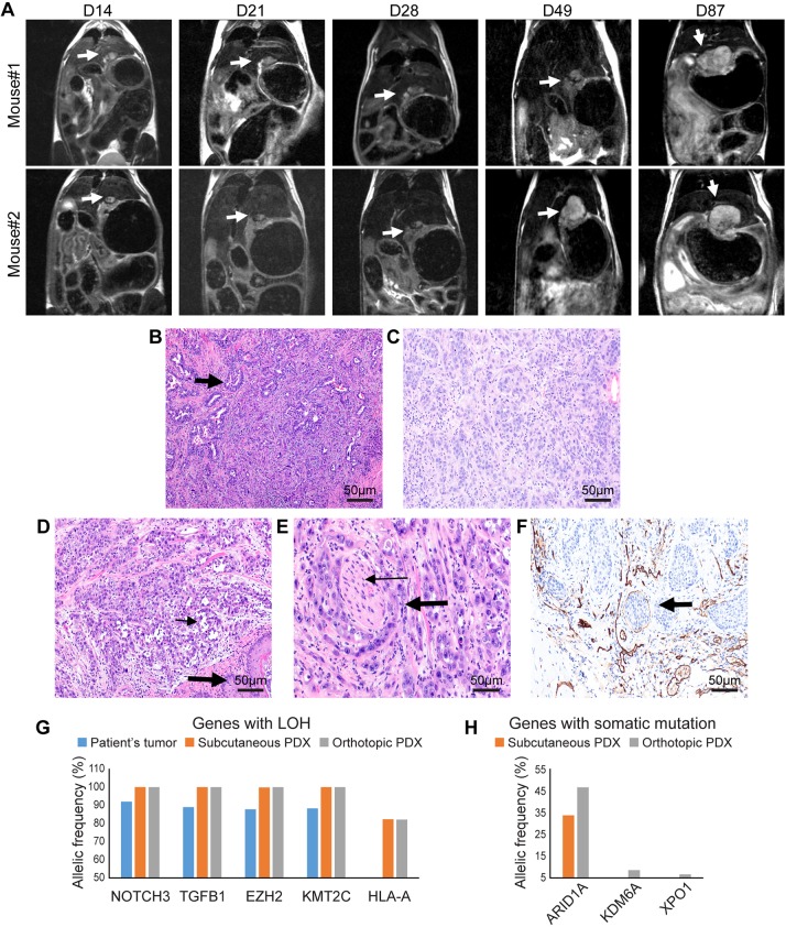
The PDOX mouse model of esophageal adenocarcinoma mimics the tumor growth pattern, and histopathological and molecular characteristics of the patient tumor. (A) MRI (coronal view) demonstrating growth of esophageal/GEJ PDOX (arrows) in two mice at day (D) 14, D21, D28, D49 and D87. In mouse #1, at D49, there is clear evidence of intra- and extraluminal spread in the esophageal wall. (B-F) Histopathological evaluation by Hematoxylin and Eosin (H&E) staining and immunohistochemistry at 200× magnification: (B) glandular architecture (arrow) and cytologic features of the patient's tumor; (C) glandular architecture and cytologic features of subcutaneously implanted PDX; (D) PDOX exhibiting glands and cellular features (thin arrow) similar to the histologic features of the patient tumor (squamous epithelium of esophagus, thick arrow); (E) a focus of perineural invasion in PDOX (nerve bundle, thin arrow; perineural tumor, thick arrow); (F) immunohistochemistry of CD31 depicting lymphovascular infiltration (arrow) in GEJ PDOX. (G,H) Next-generation sequencing results demonstrating allelic frequency (%) for genes with loss of heterozygosity (LOH) (G) and somatic mutation (H).

